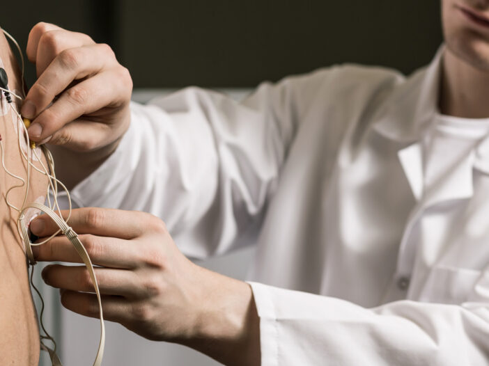
Cardiac Stress Test
A cardiac stress test measures how the heart performs in response to exercise or stress. The test monitors blood flow and oxygen levels as the heart beats faster and works harder. Stress tests are often performed to help determine the cause of chest pain or shortness-of-breath. A stress test can help diagnose coronary artery disease (CAD – narrowing of the heart’s blood vessels), which can cause chest pain and increases a patient’s risk for a heart attack. The test can also reveal irregular heart rhythms and determine if medical treatments have been effective.
Prior to the Treadmill Test, (also known as as an exercise stress test or an exercise ECG) patients are given an electrocardiograph (EKG or ECG) and an echocardiogram. The EKG measures heart rate, while the echocardiogram gives images of the heart’s structures. It assesses heart and valve health and blood flow. Both tests are painless and the patient’s heart rate, blood pressure and cardiac electrical system are monitored.
For the exercise test, electrodes are placed on the chest, arms and legs. A blood pressure cuff is worn on the arm and an oxygen monitor is placed on the finger. The patient exercises by walking on a treadmill or pedaling a bicycle until they develop symptoms or feels tired. The patient is given an EKG and echocardiogram afterward.
Metabolic stress testing – This test measures the performance of the heart and lungs while under physical stress. It is similar to an exercise stress test but includes an analysis of the patient’s respiratory system.
Pharmacological (medication-induced) stress echocardiogram – A stress echocardiogram integrates ultrasound imaging and exercise stress testing to measure how the heart functions while the patient walks on a treadmill or rides a stationary bicycle. Medication is be used to stimulate exercise for patients who are unable to exercise safely.
Nuclear stress test — Your provider may order a Nuclear Stress Test to assess the blood flow to your heart. This is done by taking two sets of pictures of your heart; one set of pictures shows the blood flow to your heart at rest, and the other set of pictures shows the blood flow to your heart at stress. Each set of pictures requires an intravenous injection of a radioactive material, which will not make you feel any different in any way. Your test may be scheduled with one set of pictures on different days, or it may be a one-day test. The stress test may be done on a treadmill, with medications, or a combination of both. For the pictures, you must lay very still on a table while a camera passes over your chest for about 15 minutes. While Nuclear Stress Testing is extremely safe, a provider will be at your side during stress testing.
Echocardiogram
A conventional or Transthoracic Echocardiogram (TTE) can be done in a resting state or during exercise (stress echo), the ultrasound source is outside the body, on top of the chest. A technician obtains views of the heart by moving a small instrument called a transducer to different locations on the chest or abdominal wall. A transducer, which resembles a microphone, sends sound waves into the chest and picks up echoes bouncing off the heart. A standard echo usually provides highly detailed images of the heart walls and chambers which can be analyzed by a cardiologist. There are no side effects from a TTE or recovery time needed.
Holter Monitor Applications and Interpretation
One tool that providers use to diagnose arrhythmia (an abnormal heart rhythm) is the Holter monitor. The Holter monitor, worn for one or two days, is a small appliance that can be attached to a belt or a shoulder strap. Several electrodes placed on the chest connect to the monitor. The monitor records heart rhythms. Patients may also be asked to keep a record of cardiac symptoms experienced while wearing the monitor. Symptoms include chest pain, heart flutters, faintness or dizziness. It is also helpful for the patient to record when he/she takes medications, exercises or experiences emotional events. The Holter monitor will not interfere with most activities, other than bathing or water-based activities, as it must remain dry.
Transesophageal Echocardiogram
Echocardiograms use sound waves bounced off the structures of the heart to generate images of the heart in motion. In some cases, most often when patients have serious lung disease, or are immobile or overweight, ultrasound images of the heart are not clear. In these cases, cardiologists may request a transesophageal echocardiography (TEE)
Unlike the standard echocardiogram, in which the transducer is placed over the chest wall, in TEE the transducer is passed into the esophagus (the swallowing tube) and sometimes into the stomach. The esophagus, in particular, provides an ideal viewing of the heart, aorta and other great vessels and produces high-quality images of these structures.
TEE examinations are useful in helping providers diagnose and evaluate patients with embolisms (clots), valvular heart disease, bacterial infections, lesions, aortic abnormalities and injuries, and congenital heart disease. TEE is also used to evaluate critically ill patients and potential heart surgery candidates. A TEE carries more risk than the standard echocardiogram procedure (which is essentially risk-free) but is still very safe and under the right circumstances can be extremely useful.
Contact Information
CMHVI Diagnostic Testing Center
60 High Street, Y1
Lewiston, Maine 04240
(207) 795-8200
Services:
- Cardiac Catheterization Lab
- Cardiac Diagnostic
- Cardiac MRI
- Cardiopulmonary Rehab
- Electrophysiology (EP) Lab
- Vascular Lab
- Vein Center
- Wound Center – 795-8260
_________________________________
Lipid Clinic
60 High Street, ground floor
Central Maine Heart Associates
(207) 753-3900
_________________________________
Single-Stay Unit (SSU)
60 High Street, Y3
Lewiston, Maine 04240
(207) 753-3907
